
In spite of its small size this foramen is used as a landmark in certain surgical procedures. The smallest area within the FIAM is occupied by the singular foramen which is located postero-inferiorly towards the inferior vestibular area, and transmits the posterior ampullar nerve.

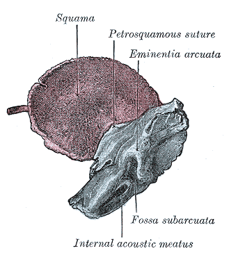
In turn, the inferior vestibular area is a place for passing the saccular nerve. The vestibular nerve originates from the superior and inferior vestibular nerves (passing through the corresponding fields within FIAM). The cochlear nerve passes through the IAM along with the facial nerve and the vestibular nerve. Superior vestibular area is a place of transition of the utriculoampullary nerve which originates from junction of the utricular nerve, anterior and lateral ampullar nerve.Ĭochlear area, situated anteriorly in the inferior part of the FIAM is a place of passage of the cochlear nerve fibres which go through the fundus of the IAM from the modiolus of the cochlea. However, this structure is not always mentioned in the papers describing morphology of the FIAM, and therefore was not included in the schematic drawing presented in Figure 1. The facial nerve area is separated from superior vestibular area by vertical osseous ridge termed as Bill’s bar which forms the vertical crest. Through the facial nerve area runs facial nerve and the intermediate nerve. The superior part of the FIAM contains: facial nerve area (situated anteriorly) and superior vestibular area (situated posteriorly), whereas the inferior part contains: cochlear area (situated anteriorly), inferior vestibular area (situated posteriorly) and singular foramen (situated postero-inferiorly). Schematic arrangement of the particular areas within the fundus of internal acoustic meatus FNA - facial nerve area SVA - superior vestibular area CA - cochlear area IVA - inferior vestibular area SF - singular foramen TC - transverse crest. Within the FIAM runs horizontally transverse crest which separates the fundus into two parts: superior and inferior, as it is presented in Figure 1.įigure 1. The FIAM transmits from the cranial cavity to ear the following structures : facial nerve, intermediate nerve, labyrinthine artery and vestibulocochlear nerve which divides near the lateral end of IAM into a two parts: a cochlear nerve and vestibular nerve. The height and width of the FIAM range from 2.5 to 4.0 mm and from 2.0 to 3.0 mm, respectively. The FIAM constitutes also the medial wall of the labyrinth. This plate separates the cochlea and vestibule from the IAM, and is defined as a fundus of the internal acoustic meatus (FIAM). Lateral end of the IAM is formed by the thin cribriform plate of bone. The entire canal has a length of about 1 cm and extends laterally inside the bone. Internal acoustic meatus (IAM) is a canal which is terminated with a fundus located inside the pyramid of the temporal bone. Key words: internal acoustic meatus, petrous bone, micro-computed tomography In the studied material we did not find out any statistically significant difference between mean diameters calculated for infant and adult individuals. Hence, we estimated the size of each area of the FIAM by measuring their minimal and maximal diameter. Application of this technique allows finding out new anatomical structures like the foramen of the transverse crest, which is not described in literature. We figured out that 3D reconstructions obtained from micro-CT scans can precisely demonstrate all areas of the FIAM (facial nerve area, cochlear area, superior and inferior vestibular areas, singular foramen). By using micro-CT we obtain detailed volume rendering images presenting topography of the FIAM in 3-dimensional (3D) space. The aim of this paper was to present micro-computed tomography (micro-CT) high resolution images of the fundus of internal acoustic meatus (FIAM) and characterise the normal appearance of its singular areas which are places of passage of numerous anatomical structures. Kopernika 12, 31–034 Kraków, Poland, e-mail: 11 September 2014 Accepted 23 November 2014] Kozerska, MSc, Department of Anatomy, Collegium Medicum, Jagiellonian University, ul. Anatomy of the fundus of the internal acoustic meatus - micro-computed tomography studyĭepartment of Anatomy, Jagiellonian University, Collegium Medicum, Krakow, PolandĪddress for correspondence: M.


 0 kommentar(er)
